NORMAL HEART RHYTHM
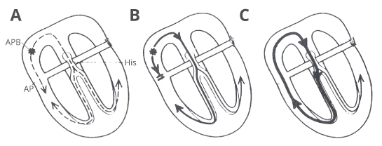
The normal heart is composed of two pumps lying side by side, each of which has a top chamber (atrium) and bottom chamber (ventricles). The heart is stimulated electrically and speeds up and slows down according to the frequency of the electrical impulses. These impulses start off from the top part of the right atrium, at a specialised bit of muscle called the sinus node. The impulse is then passed down the atrium to the junction of the atrium and the ventricles.
In the normal heart, there is a structure called the atrioventricular node, which allows impulses to conduct from atrium to ventricles. There is no other electrical connection between the atrium and the ventricles, so all impulses must pass through this atrioventricular node. These electrical impulses then pass throughout the ventricles causing them to tighten.
In some patients, there is an extra electrical connection in addition to the atrioventricular node. This is sometimes called an ‘accessory pathway’, and this focus or pathway may be causing electrical impulses to be generated or conducted wrongly. This is what causes your child’s heart to race or feel irregular.
WHAT IS AN ELECTROPHYSIOLOGY STUDY?
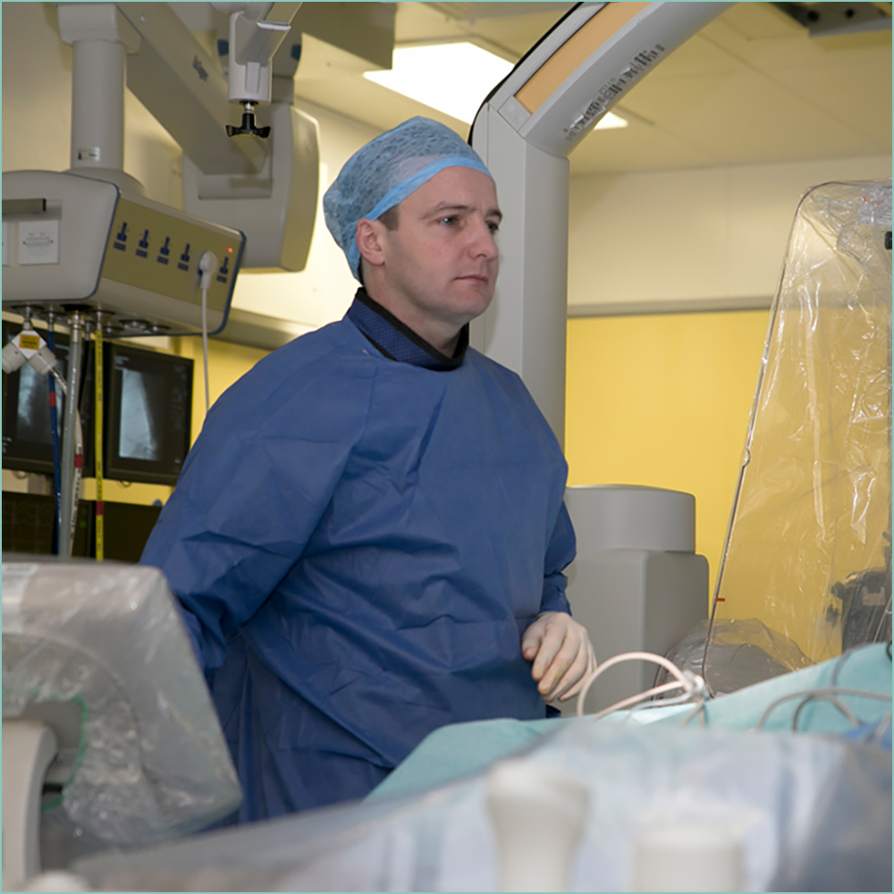
An electrophysiology study (EPS) is a procedure conducted by myself (Dr. Walsh), a heart rhythm specialist. It enables me to look at the function of the heart’s electrical system and therefore to work out the cause of your child’s strange heart rhythm. It will help the me to decide upon a course of future treatment.
WHAT IS A RADIO FREQUENCY ABLATION PROCEDURE?
It is often referred to as an ‘ablation’ procedure for short. It involves passing a small wire (called a catheter) through a vein at the top of each leg. Once the accessory pathway has been found, the catheter has heat passed through it to the tip, where it should then be possible to make a small burn (or freeze) within the heart, directly against the accessory pathway which destroys it, permanently stopping it from conducting electricity.
There are a variety of heart rhythm disorders that are treated by using the ablation process. For example, the treatment of any extra or abnormal pathways which are identified throughout the EPS process. The radio frequency ablation procedure is successful in over 95% for most patients.
WHAT HAPPENS IN THE CATHETER LAB AND HOW LONG DOES IT TAKE?
You will be put to sleep with a general anaesthetic. You will be given the opportunity to meet the consultant anaesthetist performing the procedure the morning of the procedure. Once you are asleep the catheter is inserted into the vein in the groin and moved to specific positions in the heart. Electrical conduction within the heart is monitored by sensors at the tip of these catheters. The heart is stimulated with electrical impulses and/or medicines and the catheter is manipulated within the heart. You may have EPS or EPS and ablation.
Once the procedure has finished, the catheters will be removed and you will be taken to the recovery suite.
The length of time the procedure takes will be different for each patient. It can take anywhere between 45 minutes to several hours.
HOW SOON ARE RESULTS LIKELY TO BE AVAILABLE?
I will see you after the procedure and discuss the initial results with you. On some occasions it may be necessary for you to have an echocardiogram (a scan of the heart) the day after the procedure. I will review you post procedure, and decide when you can be discharged, usually this is the day after the procedure. The recovery time is very quick and usually patients are back to all normal activity in 1-2 days. We will need to follow you up for a period of time in the cardiac outpatients.
WHAT ARE THE COMPLICATIONS?
The main risk of the procedure is the risk of damage to the atrioventricular node. The risk of injury to the atrioventricular node depends very much on where the accessory pathway is located. Probably only in about 10% of cases is there a real risk to the atrioventricular node. If the extra pathway is close to the atrioventricular node, then we tend to use a different technique called cryoablation. This essentially means freezing the pathway. This is much safer, as there is a greater degree of reversibility. Thankfully in over 1000 ablations I have never injured the atrioventricular node and needed to put a pacemaker in place.

